Onion cells Onion root cell labeled Diagram of human cheek cell and onion cell
The epidermises of onion scales. (A) Red onion bulb. B, Longitudinal
Onion epidermal drawings epidermis labeled biology chromosomes chromosome dna observation Labelled diagram of onion cell Magnified microscope cell 40x microscopy micrographs walls
Beautiful world: onion cells
Onion cell under microscope labeled[diagram] human cheek cell diagram labeled Onion cells under a microscopeOnion microscope magnified 40x 100x microscopy.
Onion micrograph epidermal cells light magnification microscopyOnion cells under microscope Onion cells microscope blue methylene stained under observation umberto flickrOnion peel cell diagram cell diagram biology art bio.

Onion root cell labeled
Onion cell peel draw cytoplasm membrane vacuole showing brainly figureOnion cell diagram drawing Onion epidermal cell diagramOnion cell peel diagram biology choose board.
The science scoop: onion cell labBiology help online: learning on onion cells Onion labeled mitosis cytokinesis interphase prophase libretexts prometaphaseOnion cell labelling diagram.

Cells cebola epiderme creativemarket containing micrograph europafotos ukphotos micrografia
Onion cells slide (80× magnification) : r/microbiologyOnion cell biology cells onions microscopic tissue structure microscopy Onion epidermal cells under microscopeOnion cell cells microscope micrograph microscopic stock skin alamy magnification root tip allium high epidermis bulb section mitosis photomicrograph show.
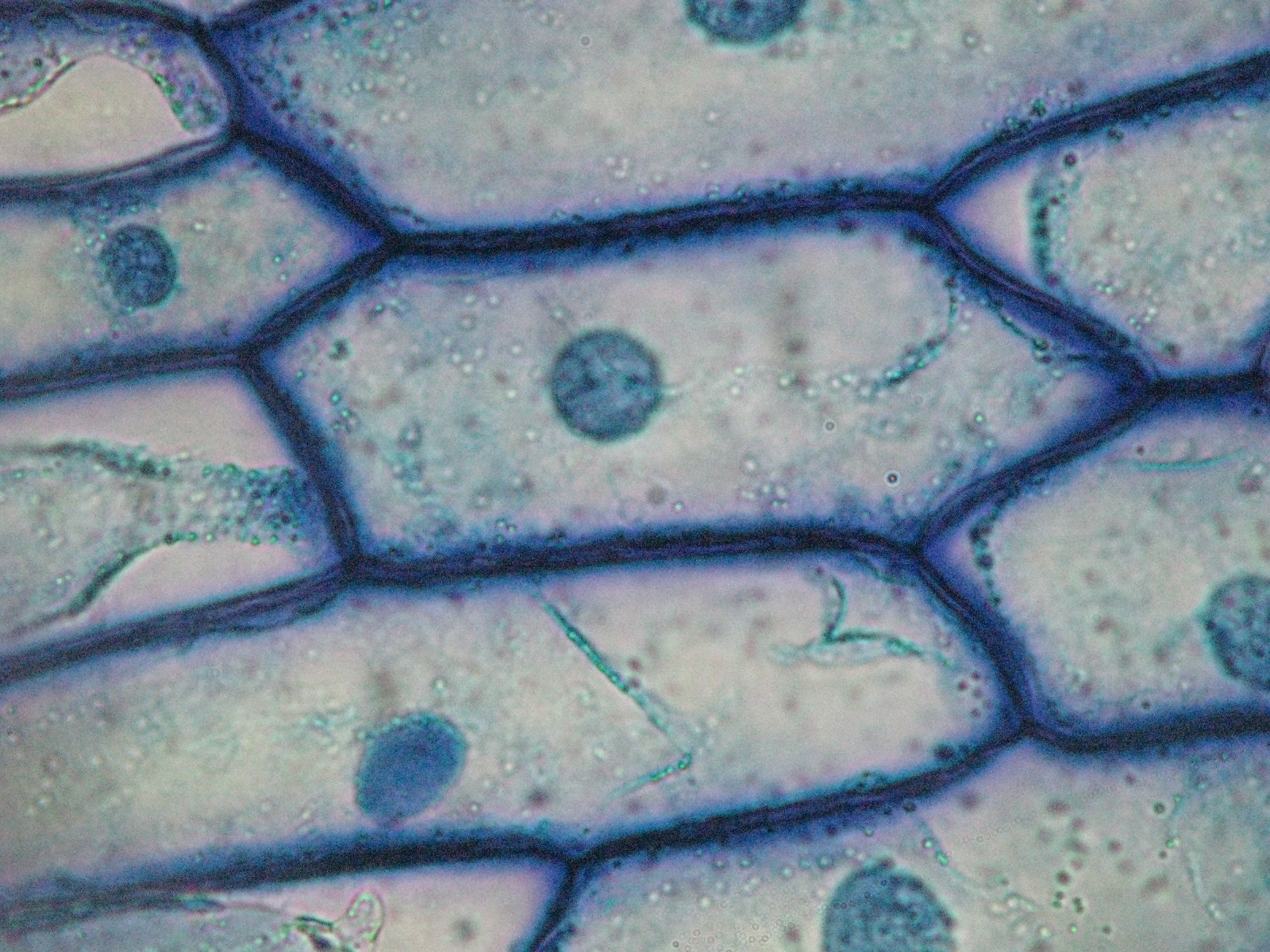
Onion cell diagram drawingOnion magnification Microscope methylene cells labelled typical epidermal biologicalRed onion; epidermal cells showing cytoplasm and nuclei; red color is.

Micrograph onion image & photo (free trial)
The epidermises of onion scales. (a) red onion bulb. b, longitudinalDraw the figure of an onion peel showing cell Onion cells beautiful world[diagram] labeled onion cell diagram.
Labeled diagram of an onion cellOnion peel cell diagram Lesson 3: onion dissection & “look at the plant cells”The layer present over the cell membrane in an onion cell is called.
![[DIAGRAM] Human Cheek Cell Diagram Labeled - MYDIAGRAM.ONLINE](https://i2.wp.com/qph.fs.quoracdn.net/main-qimg-a47ea164475ad7c915dce50ee27c8e49)
[solved] drawing of an onion cell in interface. 6. prepare a biological
Onion cell 400x lab microscope under labeled cells structure scoop science white lookedOnion cells under microscope Top 10+ what is the function of an onion to the plantOnion red cells epidermal cytoplasm color showing nuclei alamy.
Onion cell 2 biology art, cell biology, science and nature, lifeWhy are onion cells rectangular? Onion cell microscope hi-res stock photography and imagesOnion skin cell labeled diagram.

Cell onion cheek human diagram diagrams
.
.


The epidermises of onion scales. (A) Red onion bulb. B, Longitudinal

RED ONION; EPIDERMAL CELLS SHOWING CYTOPLASM AND NUCLEI; RED COLOR IS

draw the figure of an onion peel showing cell - Brainly.in
Beautiful World: Onion cells

Onion cells | High-Quality Nature Stock Photos ~ Creative Market

Lesson 3: Onion Dissection & “Look at the Plant Cells” - Rs' Science